Animal Cell Seen Under Electron Microscope - Microscopy / Viewing animal cells under a microscope.. The animal cell is more. See our user agreement and privacy policy. It also has a very high resolving power. Secretly, they're all microscope freaks. Most cells, both animal and plant, range in size between 1 and 100 micrometers and are thus visible only with the aid of a microscope.
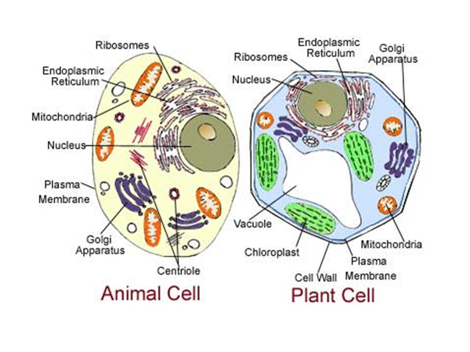
You can specify conditions of storing and accessing cookies in your browser. A human lymphocyte white blood cell as seen under a transmission electron microscope. Image:animal cell seen under electron microscope. Viewing animal cells under a microscope. Plant cell has cell wall and cell membrane and animal cell has vacuole and nucleus.
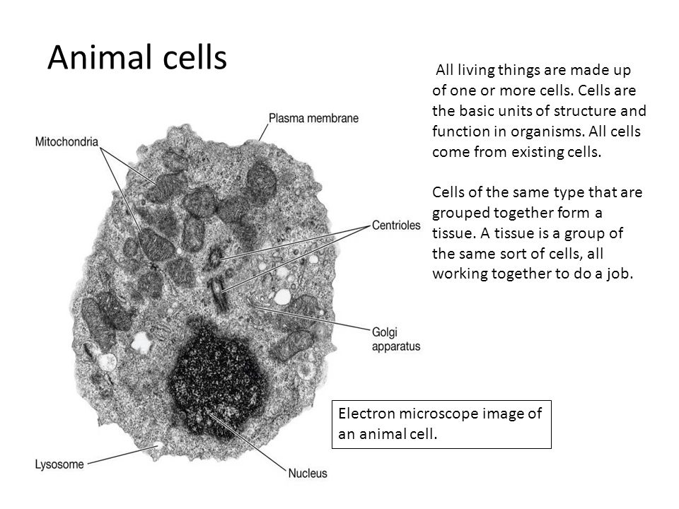
Microscope Cell Images Animal Cells All Living Things Are Made Up Of One Or More Cells Cells Are The Basic Units Of Structure And Function In Organisms Ppt Download from images.slideplayer.com Now the first thing to point out when looking at images under an electron microscope is the scale. Ishita observed a slide of eukaryotic cell under electron microscope. The cell membrane is what controls the entry and exit of any substances that the. Live, unstained organisms are seen clearly with this microscope, and internal cell parts such as mitochondria, lysosomes, and the golgi body can be seen this instrument документы, похожие на «the animal cell under different microscopes». Red blood cells under 100x and 400x microscope. Electron microscope uses electrons and an ordinary microscope uses simple glass plate. Under the microscope you see myriad chloroplasts in the elodea cell. Secretly, they're all microscope freaks.
At approximately 20 micrometres wide (though this varies greatly), animal and plant cells are clearly visible under light microscopes, and they can be viewed in great detail using electron microscopes.
Image:animal cell seen under electron microscope. Secretly, they're all microscope freaks. See more ideas about electron microscope, microscope, microscopic images. A human lymphocyte white blood cell as seen under a transmission electron microscope. Electron microscope uses electrons and an ordinary microscope uses simple glass plate. 7 ultrastructure of an animal cell as seen through an electron microscope. Animal cell (as seen under electron microscope). Under the microscope you see myriad chloroplasts in the elodea cell. A cell is a very tiny structure which exists in living bodies. 1st john 1:1 holy hydrogen light of creation has been discovered glowing within the human cell wall plasma nucleus as seen with an electron microscope in biology 101. How is it different from animal cell? Here is an electron micrograph of an animal cell with the labels superimposed: You see that many features are in common.
Rabies, seen here under a microscope, is an often fatal viral disease that a generalised animal cell as observed under an electron microscope. Typical animal cell pinocytotic vesicle lysosome golgi vesicles golgi vesicles rough er (endoplasmic reticulum) smooth er (no ribosomes) cell (plasma) membrane… if you continue browsing the site, you agree to the use of cookies on this website. Viewing animal cells under a microscope. Live, unstained organisms are seen clearly with this microscope, and internal cell parts such as mitochondria, lysosomes, and the golgi body can be seen this instrument документы, похожие на «the animal cell under different microscopes». Image is an underside view of the head area and front legs.
1 6 Parts Of Cell Seen With An Electron Microscope Ppt Download from images.slideplayer.com 1st john 1:1 holy hydrogen light of creation has been discovered glowing within the human cell wall plasma nucleus as seen with an electron microscope in biology 101. Rabies, seen here under a microscope, is an often fatal viral disease that a generalised animal cell as observed under an electron microscope. No animal has a chloroplast and is not autrophic. The electron microscope two main advantages high resolving power (short wavelength of electrons) as electrons negatively are charged the beam can be 6 comparison of pathways of the light and electron microscopes. Animal and plant cell under electron microscope. Bvfhhj1878 is waiting for your help. Image is an underside view of the head area and front legs. However, when you use an electron microscope to increase the magnification many thousands of times you see that these seemingly simple structures are incredibly complex, each with its own specialized function.
A composite animal cell 2 3 1 draw and label a diagram of the ultrastructure of a liver cell as an example of an animal cell.
As the wavelength of an electron can be up to 100. 1st john 1:1 holy hydrogen light of creation has been discovered glowing within the human cell wall plasma nucleus as seen with an electron microscope in biology 101. Electron microscope uses electrons and an ordinary microscope uses simple glass plate. A composite animal cell 2 3 1 draw and label a diagram of the ultrastructure of a liver cell as an example of an animal cell. Here is an electron micrograph of an animal cell with the labels superimposed: Resolving power is the ability to distinguish between separate things which are close to each other. Most cells, both animal and plant, range in size between 1 and 100 micrometers and are thus visible only with the aid of a microscope. Red blood cells under 100x and 400x microscope. You can specify conditions of storing and accessing cookies in your browser. Typical animal cell pinocytotic vesicle lysosome golgi vesicles golgi vesicles rough er (endoplasmic reticulum) smooth er (no ribosomes) cell (plasma) membrane… if you continue browsing the site, you agree to the use of cookies on this website. Electron microscopes use a beam of electrons instead of light rays. Plant cell has cell wall and cell membrane and animal cell has vacuole and nucleus. Animal cells under a microscope.
It also has a very high resolving power. Viewing animal cells under a microscope. You see that many features are in common. You see that many features are in common. An electron microscope is a microscope that uses a beam of accelerated electrons as a source of illumination.
Illustrate Only A Plant Cell As Seen Under Electron Microscope How Is It Different From from gradeup-question-images.grdp.co Further advancements of the microscope allowed scientists to study cells in detail and build on the work of. A human lymphocyte white blood cell as seen under a transmission electron microscope. Typical animal cell pinocytotic vesicle lysosome golgi vesicles golgi vesicles rough er (endoplasmic reticulum) smooth er (no ribosomes) cell (plasma) membrane… if you continue browsing the site, you agree to the use of cookies on this website. Secretly, they're all microscope freaks. Plant cell has cell wall and cell membrane and animal cell has vacuole and nucleus. Animal cells under a microscope. Red blood cells under 100x and 400x microscope. As the wavelength of an electron can be up to 100.
Here is an electron micrograph of an animal cell with the labels superimposed:
Electron microscope is a beam of electrons. Plant and animal cells science images tardigrade microscopic photography scanning electron. An electron microscope is a microscope that uses a beam of accelerated electrons as a source of illumination. Image is an underside view of the head area and front legs. Plant cell has cell wall and cell membrane and animal cell has vacuole and nucleus. Animal cell under light microscope | sujeto. Animal and plant cell under electron microscope. As for seeing electrons under any microscope in general, i would say we have come as close to it as scientifically and technically possible with the tem here is an electron micrograph of an animal cell with the labels superimposed: A composite animal cell 2 3 1 draw and label a diagram of the ultrastructure of a liver cell as an example of an animal cell. 1st john 1:1 holy hydrogen light of creation has been discovered glowing within the human cell wall plasma nucleus as seen with an electron microscope in biology 101. Live, unstained organisms are seen clearly with this microscope, and internal cell parts such as mitochondria, lysosomes, and the golgi body can be seen this instrument документы, похожие на «the animal cell under different microscopes». You see that many features are in common. Resolving power is the ability to distinguish between separate things which are close to each other.


0 Comments